
Magnetic Resonance Imaging
Computerized Tomography
PET/CT
Nuclear Medicine
Bone Mineral Densitometry
Click the More Info links for FAQ’s and additional information!
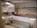
Magnetic Resonance Imaging (MRI)
Magnetic Resonance Imaging (MRI) uses a large magnet, radio waves and a computer to produce 2- or 3-dimensional images. MRI scans see right through bone and clearly picture soft tissue so they are especially valuable for helping to diagnose brain and nervous system disorders, cancer and musculoskeletal problems. MRI can also be used to diagnose other conditions and monitor the success of treatment for disease.
Our new GE System is the world’s most advanced Open MRI. It is three times as powerful as most other open systems. It combines all the advantages of a traditional high field MRI with the comfort of an open system. For the patient this means faster scanning times without sacrificing imaging quality as often occurs in most other lower field open units. With the addition of this new high field Open MRI, Central Jersey Radiologists is the only imaging center in New Jersey with three high field units. Serving the community for over 18 years, our on-site Board Certified Radiologists have more than 54 years of combined MRI experience. More info
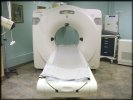
Computerized Tomography (CT)
Computerized Tomography (CT) is a special kind of x-ray study that can produce computer images of a part of the body. An x-ray tube rotates around the body scanning it with x-rays. Then a computer uses readings taken from those x-rays to make an image on a video screen. CT scans help detect conditions that regular x-ray studies can’t including tumors and blood clots. CT scans also help check progress during or after treatment. The procedure is relatively safe and painless and can often replace other diagnostic procedures such as exploratory surgery.
More info
PET/CT
What is PET/CT?
PET/CT combines the functional information from a positron emission tomography (PET) exam with the anatomical information from a computed
tomography (CT) exam into one single exam. A PET scan detects changes in cellular function – how your cells are utilizing nutrients like sugar and
oxygen. Since these functional changes take place before physical changes occur, PET can provide information that enables your physician to make an early diagnosis.
A CT scan uses a combination of X-rays and computers to give the radiologist a non-invasive way to see inside your body. One advantage of CT is its ability to rapidly acquire two-dimensional pictures of your anatomy. Using a computer these 2-D images can be presented in 3-D for in-depth clinical evaluation. The PET exam pinpoints metabolic activity in
cells and the CT exam provides an anatomical reference. When these two scans are fused together, your physician can view metabolic changes in the proper anatomical context of your body.
Why do I need this exam?
Your PET/CT exam results may have a major impact on your physician’s diagnosis of a potential health problem – and, should a disease be detected, how a treatment plan is developed and managed. A PET/CT exam not only helps your physician diagnose a problem, it also helps predict the likely outcome of various therapeutic alternatives, pinpoint the best approach to treatment, and monitor your progress. If you’re not responding as well as expected, you can be switched to a more effective therapy immediately.
What should I expect when I arrive?
When you arrive, we will take a review of your history and any past exams. For the PET portion of the exam you’ll receive a radiopharmaceutical injection. This is a radioactive tracer that must pass multiple quality control measures before it is used for any patient injection. PET radiopharmaceuticals lose their radioactivity very quickly
(2 minutes to 2 hours) and only very small amounts are injected. In all cases, little or no radioactivity will remain in your body 10 minutes to 6 hours after injection. For most studies, you’ll have to wait for the radiopharmaceutical to distribute itself – typically 30 minutes to an hour. During this time you will be asked to relax.
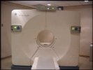
What will the scan be like?
You will lie on a comfortable padded table. The table will move
slowly through the tube-shaped PET/CT scanner as it acquires the
information needed to generate diagnostic images. You will be asked to lie very still during the scan because movement can interfere with the results. For the CT scan you will be asked to hold your breath
for a few seconds to minimize body movements. During the scan, you might hear a humming noise but you will not feel anything unusual. You may feel the table move while images are being taken at certain locations on your body. The technologist will monitor you during the exam. The specific details of your upcoming exam will be explained fully by the technologist or your physician. How long will all this take? The PET/CT scan should last between 30 and 45 minutes. The exam procedure can vary depending on what we are looking for and what we discover along
the way. Plan to spend two to three hours with us.
What happens after the exam?
You may leave us as soon as the exam is complete. Unless you’ve received special instructions, you will be able to eat and drink immediately – drinking lots of fluids soon after the exam will help remove any of the radiopharmaceutical that may still be in your system. In the meantime, we’ll begin preparing the results for review by our diagnosticians, and then
by your physician, who will tell you what we’ve learned. Safety of PET/CT exams Be assured that PET/CT exams are a safe and effective diagnostic procedure. The radiopharmaceuticals used in PET don’t remain in your system long, so there’s no reason to avoid interacting with other people once you’ve left. To be extra safe, wait for a few hours before getting too close to an infant or anyone who’s pregnant.
More info
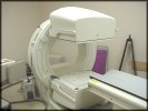
Nuclear Medicine (NM)
Nuclear medicine involves the use of small amounts of radioactive materials (tracers) to help diagnose and treat a variety of diseases. The tracer may be administered intravenously or orally, and depending on which type of scan is being performed, the imaging will be done either immediately or a few hours or even days after receiving the tracer. Procedure times vary because for many tests, a certain amount of time is needed (from a few hours to a few days) for the tracer to accumulate in the part of the body being scanned before the imaging can be done. A special camera is used that detects the tracer in the body and then records this information on a computer screen or on film.
Nuclear medicine procedures are very safe. The amount of radiation you will receive during the test is no more than what you would receive from similar x-ray procedures. The tracer you are given will remain in your body for a short period of time and is cleared from the body through natural bodily functions. Drinking plenty of fluids will help the tracer clear through your body more quickly. Adverse reactions or side effects from the tracer are rare, but let the technologist know if you experience any symptoms after the tracer is administered. For procedural information and details on specific Nuclear Medicine tests, click More info
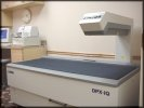
Bone Mineral Densitometry (BMD)
A bone mineral density test measures the thickness of your bones. It compares your bone measurements to two standards, known as “age matched” (your Z-score) and “young normal” (your T-score). The age-matched reading compares your bone density to what is expected in someone of your age, sex and size. The young normal reading compares your density to the optimal peak bone density of a 30 year old healthy adult of the same sex. The information is used in making a diagnosis about your bone status and fracture risk.
Bone densitometry involves a very small dose of radiation, much less than the radiation dosage of a chest x-ray. The test is painless and noninvasive.
A bone density test can detect osteoporosis before a fracture occurs and predict your chances of fractures in the future. More info
Click the More Info links for FAQ’s and additional information!